Thursday, February 24, 2011


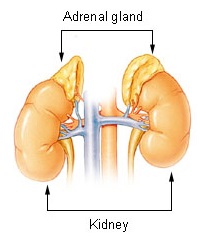
In mammals, the adrenal glands (also known as suprarenal glands) are endocrine glands that sit on top of the kidneys; in humans, the right suprarenal gland is triangular shaped while the left suprarenal gland is semilunar shaped. They are chiefly responsible for releasing hormones in conjunction with stress through the synthesis of corticosteroids such as cortisol and catecholamines, such as epinephrine. Adrenal glands affect kidney function through the secretion of aldosterone, a hormone involved in regulating plasma osmolarity.

Anatomy and function

Anatomically, the adrenal glands are located in the retroperitoneum situated atop the kidneys, one on each side. They are surrounded by an adipose capsule and renal fascia. In humans, the adrenal glands are found at the level of the 12th thoracic vertebra. Each adrenal gland is separated into two distinct structures, the adrenal cortex and medulla, both of which produce hormones. The cortex mainly produces cortisol, aldosterone, and androgens, while the medulla chiefly produces epinephrine and norepinephrine. The combined weight of the adrenal glands in an adult human ranges from 7 to 10 grams.

Cortex

The adrenal cortex is devoted to the synthesis of corticosteroid hormones. Specific cortical cells produce particular hormones including cortisol, corticosterone, androgens such as testosterone, and aldosterone. Under normal unstressed conditions, the human adrenal glands produce the equivalent of 35–40 mg of cortisone acetate per day.[2] In contrast to the direct innervation of the medulla, the cortex is regulated by neuroendocrine hormones secreted by the pituitary gland and hypothalamus, as well as by the renin-angiotensin system.

The adrenal cortex comprises three zones, or layers. This anatomic zonation can be appreciated at the microscopic level, where each zone can be recognized and distinguished from one another based on structural and anatomic characteristics. The adrenal cortex exhibits functional zonation as well: by virtue of the characteristic enzymes present in each zone, the zones produce and secrete distinct hormones.

Zona glomerulosa (outer)
The outermost layer, the zona glomerulosa is the main site for production of mineralocorticoids, mainly aldosterone, which is largely responsible for the long-term regulation of blood pressure.
Zona fasciculata
Situated between the glomerulosa and reticularis, the zona fasciculata is responsible for producing glucocorticoids, chiefly cortisol in humans. The zona fasciculata secretes a basal level of cortisol but can also produce bursts of the hormone in response to adrenocorticotropic hormone (ACTH) from the anterior pituitary.
Zona reticularis
The inner most cortical layer, the zona reticularis produces androgens, mainly dehydroepiandrosterone (DHEA) and DHEA sulfate (DHEA-S) in humans.Medulla

The adrenal medulla is the core of the adrenal gland, and is surrounded by the adrenal cortex. The chromaffin cells of the medulla, named for their characteristic brown staining with chromic acid salts, are the body's main source of the circulating catecholamines adrenaline (epinephrine) and noradrenaline (norepinephrine). Derived from the amino acid tyrosine, these water-soluble hormones are major hormones underlying the fight-or-flight response.

To carry out its part of this response, the adrenal medulla receives input from the sympathetic nervous system through preganglionic fibers originating in the thoracic spinal cord from T5–T11.[4] Because it is innervated by preganglionic nerve fibers, the adrenal medulla can be considered as a specialized sympathetic ganglion.[4] Unlike other sympathetic ganglia, however, the adrenal medulla lacks distinct synapses and releases its secretions directly into the blood.

Cortisol also promotes epinephrin synthesis in the medulla. Produced in the cortex, cortisol reaches the adrenal medulla and at high levels, the hormone can promote the upregulation of phenylethanolamine N-methyltransferase (PNMT), thereby increasing epinephrine synthesis and secretion.

Blood supply

Although variations of the blood supply to the adrenal glands (and indeed the kidneys themselves) are common, there are usually three arteries that supply each adrenal gland:

Venous drainage of the adrenal glands is achieved via the suprarenal veins:

The suprarenal veins may form anastomoses with the inferior phrenic veins.

The adrenal glands and the thyroid gland are the organs that have the greatest blood supply per gram of tissue. Up to 60 arterioles may enter each adrenal gland. This may be one of the reasons lung cancer commonly metastasizes to the adrenals.
Pineal Gland


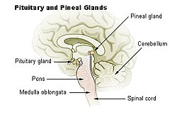
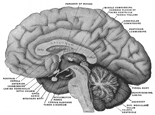
The pineal gland (also called the pineal body, epiphysis cerebri, epiphysis or the "third eye") is a small endocrine gland in the vertebrate brain. It produces the serotonin derivative melatonin, a hormone that affects the modulation of wake/sleep patterns and seasonal functions.[1][2] Its shape resembles a tiny pine cone (hence its name), and it is located near the center of the brain, between the two hemispheres, tucked in a groove where the two rounded thalamic bodies join.

Function

The pineal gland was originally believed to be a "vestigial remnant" of a larger organ. In 1917 it was known that extract of cow pineals lightened frog skin. Dermatology professor Aaron B. Lerner and colleagues at Yale University, hoping that a substance from the pineal might be useful in treating skin diseases, isolated and named the hormone melatonin in 1958.[13] The substance did not prove to be helpful as intended, but its discovery helped solve several mysteries such as why removing the rat's pineal accelerated ovary growth, why keeping rats in constant light decreased the weight of their pineals, and why pinealectomy and constant light affect ovary growth to an equal extent; this knowledge gave a boost to the then new field of chronobiology.[14]

Melatonin is N-acetyl-5-methoxy-tryptamine, a derivative of the amino acid tryptophan, which also has other functions in the central nervous system. The production of melatonin by the pineal gland is stimulated by darkness and inhibited by light.[15] Photosensitive cells in the retina detect light and directly signal the SCN, entraining its rhythm to the 24-hour cycle in nature. Fibers project from the SCN to the paraventricular nuclei (PVN), which relay the circadian signals to the spinal cord and out via the sympathetic system to superior cervical ganglia (SCG), and from there into the pineal gland. The function(s) of melatonin in humans is not clear; it is commonly prescribed for the treatment of circadian rhythm sleep disorders.

The compound pinoline is also produced in the pineal gland; it is one of the beta-carbolines.

The human pineal gland grows in size until about 1–2 years of age, remaining stable thereafter,[16][17] although its weight increases gradually from puberty onwards.[18][19] The abundant melatonin levels in children are believed to inhibit sexual development, and pineal tumors have been linked with precocious puberty. When puberty arrives, melatonin production is reduced. Calcification of the pineal gland is typical in adults.

Pituitary Gland


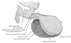
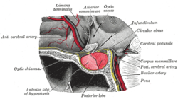
In vertebrate anatomy the pituitary gland, or hypophysis, is an endocrine gland about the size of a pea and weighing 0.5 g (0.02 oz.), in humans. It is a protrusion off the bottom of the hypothalamus at the base of the brain, and rests in a small, bony cavity (sella turcica) covered by a dural fold (diaphragma sellae). The pituitary is functionally connected to the hypothalamus by the median eminence via a small tube called the infundibular stem (Pituitary Stalk). The pituitary fossa, in which the pituitary gland sits, is situated in the sphenoid bone in the middle cranial fossa at the base of the brain. The pituitary gland secretes nine hormones that regulate homeostasis.

Hormones secreted from the pituitary gland help control the following body processes:

Tuesday, February 15, 2011

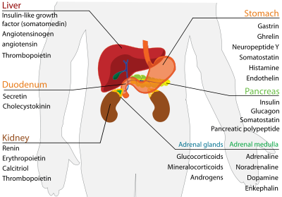
Endocrine organs and secreted hormones

Diseases of the endocrine system

Diseases of the endocrine system are common,[14] including conditions such as diabetes mellitus, thyroid disease, and obesity. Endocrine disease is characterized by disregulated hormone release (a productive pituitary adenoma), inappropriate response to signaling (hypothyroidism), lack of a gland (diabetes mellitus type 1, diminished erythropoiesis in chronic renal failure), or structural enlargement in a critical site such as the thyroid (toxic multinodular goitre). Hypofunction of endocrine glands can occur as a result of loss of reserve, hyposecretion, agenesis, atrophy, or active destruction. Hyperfunction can occur as a result of hypersecretion, loss of suppression, hyperplastic or neoplastic change, or hyperstimulation.

Endocrinopathies are classified as primary, secondary, or tertiary. Primary endocrine disease inhibits the action of downstream glands. Secondary endocrine disease is indicative of a problem with the pituitary gland. Tertiary endocrine disease is associated with dysfunction of the hypothalamus and its releasing hormones.[citation needed]

As the thyroid, and hormones have been implicated in signaling distant tissues to proliferate, for example, the estrogen receptor has been shown to be involved in certain breast cancers. Endocrine, paracrine, and autocrine signaling have all been implicated in proliferation, one of the required steps of oncogenesis.

Endocrine organs and secreted hormones

Endocrine System


In physiology, the endocrine system is a system of glands, each of which secretes a type of hormone into the bloodstream to regulate the body. It derives from the Greek words endo (Greek ένδο) meaning inside, within, and crinis (Greek κρινής) for secrete. The endocrine system is an information signal system like the nervous system. Hormones are substances (chemical mediators) released from endocrine tissue into the bloodstream that attach to target tissue and allow communication among cells. Hormones regulate many functions of an organism, including mood, growth and development, tissue function, and metabolism. The field of study that deals with disorders of endocrine glands is endocrinology, a branch of internal medicine.


The endocrine system is made up of a series of ductless glands that produce chemicals called hormones. A number of glands that signal each other in sequence is usually referred to as an axis, for example, the hypothalamic-pituitary-adrenal axis. Typical endocrine glands are the pituitary, thyroid, and adrenal glands. Features of endocrine glands are, in general, their ductless nature, their vascularity, and usually the presence of intracellular vacuoles or granules storing their hormones. In contrast, exocrine glands, such as salivary glands, sweat glands, and glands within the gastrointestinal tract, tend to be much less vascular and have ducts or a hollow lumen.

In addition to the specialised endocrine organs mentioned above, many other organs that are part of other body systems, such as the kidney, liver, heart and gonads, have secondary endocrine functions. For example the kidney secretes endocrine hormones such as erythropoietin and renin.

Struktur Neuron

Sistem Saraf Autonomi

Neurotransmission


Neurotransmission (Latin: transmissio = passage, crossing; from transmitto = send, let through), also called synaptic transmission, is an electrical movement within synapses caused by a propagation of nerve impulses. As each nerve cell receives neurotransmitter from the presynaptic neuron, or terminal button, to the postsynaptic neuron, or dendrite, of the second neuron, it sends it back out to several neurons, and they do the same, thus creating a wave of energy until the pulse has made its way across an organ or specific area of neurons.

Nerve impulses are essential for the propagation of signals. These signals are sent to and from the central nervous system via efferent and afferent neurons in order to coordinate smooth, skeletal and cardiac muscles, bodily secretions and organ functions critical for the long-term survival of multicellular vertebrate organisms such as mammals.

Neurons form networks through which nerve impulses travel. Each neuron receives as many as 15,000 connections from other neurons. Neurons do not touch each other; they have contact points called synapses. A neuron transports its information by way of a nerve impulse. When a nerve impulse arrives at the synapse, it releases neurotransmitters, which influence another cell, either in an inhibitory way or in an excitatory way. The next neuron may be connected to many more neurons, and if the total of excitatory influences is more than the inhibitory influences, it will also "fire", that is, it will create a new action potential at its axon hillock, in this way passing on the information to yet another next neuron, or resulting in an experience or an action.

CASPER


Reka Letak
Reka letak dalam perisian persembahan elektronik ialah ilmu atau kemahiran yang digunakan untuk meletak dan menyusun media pada paparan bagi membentuk persembahan yang menarik. Keupayaan meletak dan menyusun elemen media yang baik dan berkesan memberi kesan yang positif terhadap persembahan.


Prinsip Reka Letak Casper
Prinsip reka letak CASPER merupakan panduan menyusun media seperti teks, grafik, animasi dan video pada sesuatu persembahan yang dapat merangsang perhatian dan minat penonton. Skrin yang diletakan dengan media tanpa di rancangan menyebabkan persembahan yang kurang menarik.

(i) Contrast (Pebezaan yang Ketara)
Bagi menunjukkan perbezaan antara dua objek, pembangun perisian perlulah memilih warna yang kontra (warna gelap dengan warna terang). Kontra juga digunakan untuk membezakan antara unsur dengan latar belakang paparan. Selain daripada membezakan dua item yang berlainan, kontra adalah salah satu cara yang paling berkesan untuk menambahkan tarikan atau perhatian terhadap sesuatu reka bentuk. Selain daripada perbezaan warna, kontra juga boleh ditonjolkan dengan perbezaan font, objek besar dan kecil, garis halus dengan kasar, tekstur licin dengan kasar, unsur mendatar dengan menegak, garis berjauhan dengan berdekatan dan grafik kecil dengan besar.

(ii) Alignment (Susunan Lurus)
Dalam prinsip alignment, setiap item yang hendak diletakkan dalam muka perisian perlulah seimbang dengan muka perisian agar kelihatan menarik. Pembangun perisian seharusnya pandai menyusun item-item yang ingin dipersembahkan supaya ia kelihatan seimbang. Dengan kata lain, dalam prinsip Alignment, item perlu disusun supaya tidak janggal. Item juga perlu mempunyai kaitan secara visual antara satu sama lain di setiap paparan perisian.

(iii) Simplicity (Mudah)
Grafik dan animasi perlu ringkas dan dapat merangsangkan pengguna memahami maksud yang hendak disampaikan. Dengan kata lain sesuatu bahan grafik dan bahan yang diletakkan di atas muka perisian seharusnya dapat memudahkan penerokaan dan merangsangkan pemikiran. Visual yang dipilih perlulah ringkas, bersesuaian dengan pengguna dan mudah difahami. Dengan kata lain, penggunaan grafik dan elemen media dalam perisian yang dibina perlulah membantu pengguna melayari perisian dengan mudah. Sekeping gambar yang ringkas contohnya, lebih bermakna dari teks yang panjang lebar.

(iv) Proximity (Berhampiran)
Dalam prinsip proximity item yang digunakan dalam muka perisian perlulah dikumpulkan pada satu kawasan, supaya pengguna dapat melihat kesinambungan yang wujud antara item. Pengumpulan item membuatkan pengguna merasa selesa kerana item-item yang berkaitan dilihat sebagai satu kumpulan dan tidak terpisah-pisah. Oleh yang demikian, elemen ini
akan menjadi satu unit visual daripada beberapa unit terpisah.

(v) Emphasis (Penekanan)
Pembangun perisian perlu menggunakan cara tertentu bagi memberi penekanan terhadap perkara yang dirasakan penting. Pelbagai cara digunakan oleh pembangun perisian untuk menarik perhatian pengguna. Namun begitu adalah dinasihati supaya pembangun perisian tidak
menggunakan terlalu banyak elemen sampingan untuk menarik perhatian pengguna, yang mana akan menyebabkan tumpuan pengguna lebih kepada elemen sampingan.

(vi) Repetition (Pengulangan)
Dalam prinsip repetition terdapat satu piawai antara muka perisian supaya pengguna akan dapat mentafsir dan memahami dengan mudah arahan yang diberikan oleh perisian. Pengulangan menggunakan persembahan media yang berlainan akan membantu pemahaman pengguna. Ini adalah kerana pengguna perisian terdiri daripada mereka yang mempunyai
kecenderungan yang terhadap media.Pembangun perisian boleh menggunakan perulangan media teks, audio, grafik dan video untuk menyampaikan mesej yang sama.

Pengaliran Impuls Saraf

Neuron Perantaraan

Neuron Deria & Motor

Sunday, February 13, 2011

Sympathetic & Parasympathetic


The Sympathetic Nervous System

The preganglionic motor neurons of the sympathetic system (shown in black) arise in the spinal cord. They pass into sympathetic ganglia which are organized into two chains that run parallel to and on either side of the spinal cord.

The preganglionic neuron may do one of three things in the sympathetic ganglion:
  • synapse with postganglionic neurons (shown in white) which then reenter the spinal nerve and ultimately pass out to the sweat glands and the walls of blood vessels near the surface of the body.
  • pass up or down the sympathetic chain and finally synapse with postganglionic neurons in a higher or lower ganglion
  • leave the ganglion by way of a cord leading to special ganglia (e.g. the solar plexus) in the viscera. Here it may synapse with postganglionic sympathetic neurons running to the smooth muscular walls of the viscera. However, some of these preganglionic neurons pass right on through this second ganglion and into the adrenal medulla. Here they synapse with the highly-modified postganglionic cells that make up the secretory portion of the adrenal medulla.

The neurotransmitter of the preganglionic sympathetic neurons is acetylcholine (ACh). It stimulates action potentials in the postganglionic neurons.

The neurotransmitter released by the postganglionic neurons is noradrenaline (also called norepinephrine).

The action of noradrenaline on a particular gland or muscle is excitatory is some cases, inhibitory in others. (At excitatory terminals, ATP may be released along with noradrenaline.)

The release of noradrenaline
  • stimulates heartbeat
  • raises blood pressure
  • dilates the pupils
  • dilates the trachea and bronchi
  • stimulates the conversion of liver glycogen into glucose
  • shunts blood away from the skin and viscera to the skeletal muscles, brain, and heart
  • inhibits peristalsis in the gastrointestinal (GI) tract
  • inhibits contraction of the bladder and rectum
  • and, at least in rats and mice, increases the number of AMPA receptors in the hippocampus and thus increases long-term potentiation (LTP).

In short, stimulation of the sympathetic branch of the autonomic nervous system prepares the body for emergencies: for "fight or flight" (and, perhaps, enhances the memory of the event that triggered the response).

Activation of the sympathetic system is quite general because
  • a single preganglionic neuron usually synapses with many postganglionic neurons;
  • the release of adrenaline from the adrenal medulla into the blood ensures that all the cells of the body will be exposed to sympathetic stimulation even if no postganglionic neurons reach them directly.

The Parasympathetic Nervous System

The main nerves of the parasympathetic system are the tenth cranial nerves, the vagus nerves. They originate in the medulla oblongata. Other preganglionic parasympathetic neurons also extend from the brain as well as from the lower tip of the spinal cord.

Each preganglionic parasympathetic neuron synapses with just a few postganglionic neurons, which are located near — or in — the effector organ, a muscle or gland. Acetylcholine (ACh) is the neurotransmitter at all the pre- and many of the postganglionic neurons of the parasympathetic system. However, some of the postganglionic neurons release nitric oxide (NO) as their neurotransmitter.


The Nobel Prize winning physiologist Otto Loewi discovered (in 1920) that the effect of both sympathetic and parasympathetic stimulation is mediated by released chemicals. He removed the living heart from a frog with its sympathetic and parasympathetic nerve supply intact. As expected, stimulation of the first speeded up the heart while stimulation of the second slowed it down.

Loewi found that these two responses would occur in a second frog heart supplied with a salt solution taken from the stimulated heart. Electrical stimulation of the vagus nerve leading to the first heart not only slowed its beat but, a short time later, slowed that of the second heart also. The substance responsible was later shown to be acetylcholine. During sympathetic stimulation, adrenaline (in the frog) is released.

Parasympathetic stimulation causes
  • slowing down of the heartbeat (as Loewi demonstrated)
  • lowering of blood pressure
  • constriction of the pupils
  • increased blood flow to the skin and viscera
  • peristalsis of the GI tract

In short, the parasympathetic system returns the body functions to normal after they have been altered by sympathetic stimulation. In times of danger, the sympathetic system prepares the body for violent activity. The parasympathetic system reverses these changes when the danger is over.

The vagus nerves also help keep inflammation under control. Inflammation stimulates nearby sensory neurons of the vagus. When these nerve impulses reach the medulla oblongata, they are relayed back along motor fibers to the inflamed area. The acetylcholine from the motor neurons suppresses the release of inflammatory cytokines, e.g., tumor necrosis factor (TNF), from macrophages in the inflamed tissue.

Although the autonomic nervous system is considered to be involuntary, this is not entirely true. A certain amount of conscious control can be exerted over it as has long been demonstrated by practitioners of Yoga and Zen Buddhism. During their periods of meditation, these people are clearly able to alter a number of autonomic functions including heart rate and the rate of oxygen consumption. These changes are not simply a reflection of decreased physical activity because they exceed the amount of change occurring during sleep or hypnosis.

Reflex Arc

Skeletal System

Planes of the Body

Body Orientation and Direction

Fungsi Sistem Saraf


Patellar reflex: when the patellar tendon is tapped just below the knee, the patellar reflex is initiated and the lower leg kicks forward (via contraction of the quadriceps). The tap initiates an action potential in a specialised structure known as a muscle spindle located within the quadriceps. This action potential travels to the spinal cord, via a sensory axon which chemically communicates by releasing glutamate (see synapse) onto a motor nerve. The result of this motor nerve activity is contraction of the quadriceps muscle, leading to extension of the lower leg at the knee. The sensory input from the quadriceps also activates local interneurons that release the inhibitory neurotransmitter glycine onto motor neurons, blocking the innervation of the antagonistic (hamstring) muscle. The relaxation of the opposing muscle facilitates extension of the lower leg.


Central Nervous System

Otot Rangka (Pandangan Belakang)

Otot Rangka

Fungsi Otot

Sliding Filament